
Scientists produce X-rays by accelerating electrons (generally “boiled” off a heated filament) through a large potential difference and colliding these high-energy electrons into a metal target, such as molybdenum, platinum, or tungsten. An evacuated glass tube generally houses the heated filament and the metal target. As energetic electrons bombard the metal target, two basic types of X-rays appear in a plot of X-ray intensity per unit wavelength versus wavelength. First are the sharp peaks called characteristic X-rays—distinctive lines superimposed upon a broad spectrum that are peculiar to and characteristic of the target material. Scientists call the broad continuous spectrum that also appears bremsstrahlung (German for “braking radiation”). As electrons decelerate, or “brake,” upon hitting a target material, they give up energy by emitting bremsstrahlung X-rays. The field of medical radiology emerged almost immediately after Roentgen’s discovery of X-rays in 1895.
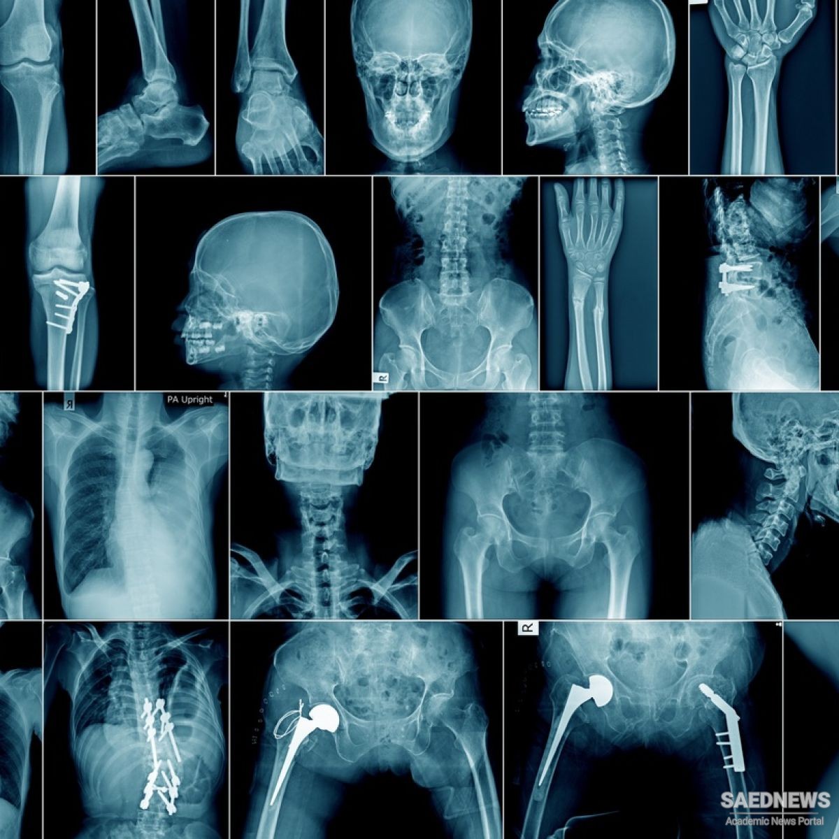
In conventional radiography, the radiologist positions the patient in front of a special photographic film, which is sensitive (that is, it darkens) when exposed to X-rays. The health worker then sends a beam of X-rays through the patient’s body. As the X-rays pass through the patient’s body, bone and other dense tissue absorb the ionizing radiation. Consequently, the photographic film behind the patient’s bones is less exposed and a pale skeletal shadow appears. Soft tissue produces an intermediate gray tone, while less-dense body organs, such as the air-filled lungs, allow the X-rays to pass through relatively unimpeded, causing the corresponding region of the photographic film to become significantly exposed and appear black when the film is developed. Contemporary radiographic film often contains radiation-sensitive photographic film enhanced by a layer of fluorescent material that glows when exposed to X-rays. This additional optical stimulation increases the contrast of the exposed film and results in a medical X-ray image of much greater diagnostic value.
 Nuclear Technology and New Ways of Tomography